When one of our consultants collects samples for asbestos identification on a client’s property, we are frequently asked if we can tell whether the material is positive or not for asbestos, and our typical answer is: “It is hard to say, only after testing can we know for sure”. Well, IT IS REALLY HARD to have any conclusion by the “naked eye” and to confirm the presence or absence of asbestos, we need to analyse the material under the microscope . So, to the curious people that would like to have an idea of what happens when we receive a sample to be tested in our laboratory, we will try to explain in a short and simple way how the identification of asbestos in building materials is done.
First, we need to know that all types of asbestos are anisotropic minerals, meaning that the speed of light varies depending on the direction through the mineral, which implies different optical properties in different directions of the fibres when viewing in a polarised light. The physical (e.g., appearance, size, and colours) and optical characteristics under a Stereo Microscope and Polarised Light Microscope (PLM) are very well defined in literature, so the test consists of observing the fibres under both microscopes, and when we have a combination of the physical and optical characteristics present, we can confirm the presence (and type) or absence of asbestos.
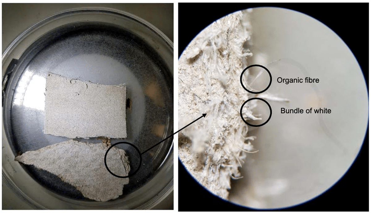
When we receive a sample in the lab, we observe it under the Stereo Microscope, with a magnification up to 40x, looking for the presence of any type of fibre on the surface and inside the material (including different layers). Figure 1 is an example of one of the most common materials received in the lab – a piece of cement board, and Figure 2 is the same piece of cement board but magnified 40 times.

Figure 1. Piece of a broken cement board as received in lab for analysis.
Figure 2. Same piece of cement board as figure 1, observing the edge of the board with 40x magnification under stereo microscope.
The method most accepted for asbestos ID analysis in New Zealand is in accordance with the Australian Standard AS4964:2004 Method for the Qualitative Identification of Asbestos in Bulk Samples using ‘Polarised Light Microscope’ including ‘Dispersion Staining Technique’.
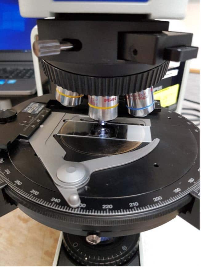
There are 6 types of asbestos – the most common (most used commercially) are white (chrysotile), brown (amosite) and blue (crocidolite) asbestos.The asbestos analyst needs a lot of training and experience to be able to differentiate all types of asbestos from other fibres that can be present in the material, such as organic fibres and fibre glass. After we have observed the presence of fibres, we will mount the observed fibres on a microscope slide with the appropriate Refractive Index (RI) Liquid (for each type of asbestos fibres, there is a specific liquid) and observe those fibres under the PLM with 100x magnification (Figure 3).

Figure 3. White asbestos fibres mounted on a slide using 1.55 RI liquid and ready to be analysed using PLM.
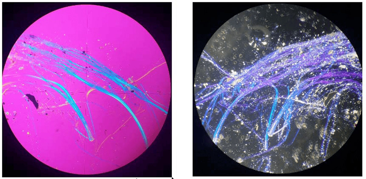
On the PLM, the analyst will observe the change in colours according to the direction of the fibre and the polarised light. Using different filters and microscope settings, the analyst will observe several characteristics, including Asbestos Form, Sign of Elongation (Figure 4), Birefringence, Extinction Angle, Pleochroism and Dispersion Staining (Figure 5). If the fibres observed have most or all the expected characteristics, including the right colours for Dispersion Staining, the analyst will be able to confirm if the fibres are related to asbestos (and the type) or not.


